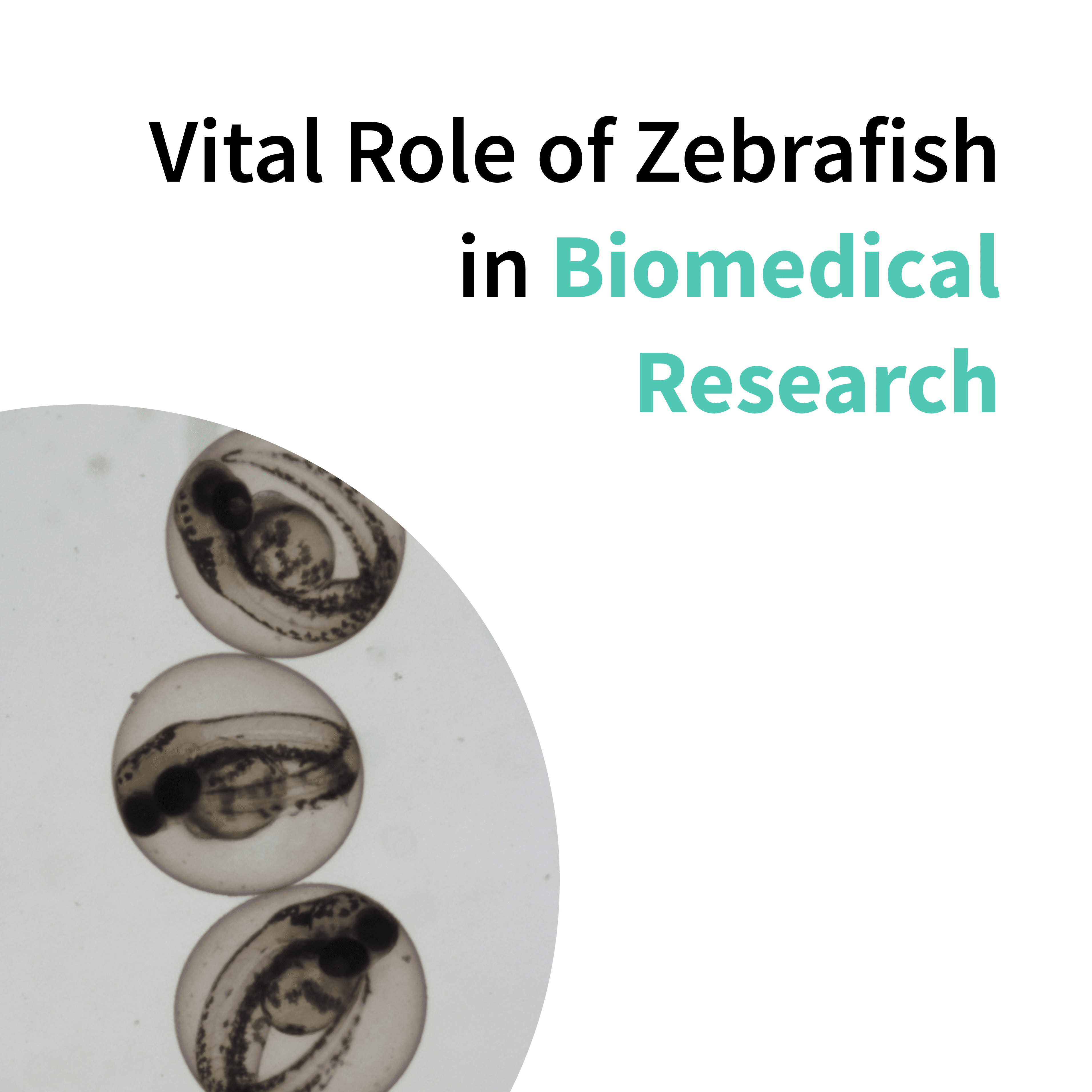
Vital Role of Zebrafish in Biomedical Research
The application of animal models in research has significantly advanced our understanding of complex biological processes and disease mechanisms. Among different animal models, rodents are the most common model in biomedical research. However, alternative models such as Zebrafish lately have been getting greater recognition in developmental biology, especially in the study of developmental stages of vertebrates.
In this article, we will elucidate the vital applications of Zebrafish in research and its importance in biomedical research, reviewing all the Zebrafish developmental stages.
Zebrafish Development in Biomedical Research
Zebrafish research has grown exponentially since it was first introduced as a model organism in the 1960s. Many key features support the use of zebrafish in scientific studies over rodent models. These include Zebrafish external embryonic development, outside of the mother’s body, hundreds of embryos growing in a single clutch, and optical clarity of the developing organism, which allows live imaging and phenotypical screening at the organism level. Moreover, the high similarity of their organs to mammals, plus the ease with which they can be genetically modified, make them a leading model for vertebrate development and disease modeling studies.
However, the fast organogenesis is the most attractive feature of using Zebrafish in scientific studies as a NAM (New Alternative Model) in development research. Most of the organs of Zebrafish develop within hours post-fertilization (hpf), as opposed to other animal models, which take days to develop fully. Their rapid life cycle progresses from fertilization to a fully formed organism in 3 days. Moreover, each stage of Zebrafish development can be exploited for different applications.
We will outline each stage of Zebrafish embryonic development and the specific applications suitable for each phase.
Key Applications: Developmental Stages of Zebrafish in Scientific Studies
Stage 1: Zygote
Zebrafish embryonic development begins with the fertilization, proceeding through a series of rapid and synchronous cell divisions known as cleavages. These divisions do not increase the embryo's size but partition the zygote into smaller cells called blastomeres. Approximately in 45 minutes post-fertilization, the first cleavage occurs, initiating the zygote stage. This phase is crucial for studies on cell division mechanisms and the early determination of cell fate.
Stage 2: Blastula
Zebrafish embryonic development follows toward the blastula phase. During this stage, cell divisions continue, become asynchronous, and the embryo reaches about 1000 cells in just 3 hpf. The blastula expands as cells start to differentiate slightly. A key feature of this period is the blastocoel formation, a fluid-filled cavity within the embryo. The activation of zygotic gene expression marks the mid-blastula transition.
Zebrafish research is critical in this period, especially for understanding early developmental signaling and gene regulation mechanisms. Moreover, acute toxicity studies can be initiated as early as 2 hours post-fertilization, such as the Teratotox Assay developed by Biobide, which can be performed to evaluate the teratogenic potential of a compound of interest during the whole development process, therefore starting from 2-4 hpf until 4 to 5 dpf.
Stage 3: Gastrulation
Gastrulation, happening around 5.5 to 10 hpf, is a crucial stage of Zebrafish embryonic development, where the embryo reorganizes from a simple blastula to a multi-layered structure. This process establishes the three primary germ layers: ectoderm, mesoderm, and endoderm. Cell movements are extensive during gastrulation, including involution, convergence, and extension, shaping the embryonic body plan. Gastrulation is fundamental for studying cell migration, differentiation, and the early establishment of the body axis.

Stages of embryonic development of the Zebrafish. Adapted from Kimmel et al. Stages of embryonic development of zebrafish. Dev. Dyn. 1995;203:253-310.
Stage 4: Segmentation
The segmentation stage, somitogenesis, extends from approximately 10 to 24 hpf. During this time, the notochord and somites—blocks of mesodermal tissue that will give rise to vertebrae and associated muscles—are formed. Zebrafish begin to exhibit the segmented organization characteristic of vertebrates. Additionally, the rudimentary nervous system starts to take shape. The segmentation period is vital for research into musculoskeletal development, neurogenesis, and the establishment of the body's segmented architecture. Somite alteration is one of the most representative morphological alterations related to chemical developmental toxicity, making this stage suitable for developing phenotypic toxicity studies based on morphologic alterations.
Stage 5: Pharyngula
The pharyngula stage starts around 24 to 48 hpf when the pharyngeal arches become prominent. It is characterized by a rapid growth and differentiation. For this reason, starting from this developmental stage, Zebrafish can be used to carry out organ-specific assays. In this phase, the heart and the circulatory system begin to work, and this characteristic is exploited in assays such as the Cardiotox Assay of novel chemical compounds. Biobide has developed the Cardiotox Assay to evaluate the effects of a compound of interest on different cardiac dysfunctions, such as arrhythmias, bradycardia, cardiac arrest, or heart failure. Key features such as the brain, spinal cord, and other organs continue to develop and mature. The pharyngula period is crucial for studying organogenesis, neural development, and the effects of genetic mutations or environmental factors on these processes.
Stage 6: Hatching
The hatching stage occurs between 48 and 72 hpf, culminating in the embryo being released from the protective chorion. An increased activity of the developing zebrafish and the beginning of the exogenous feeding marks this stage.
Before 5-6 days post-fertilization (dpf), they are not still considered animals under experimental regulatory definitions. For this reason, they can be exploited as an advantageous NAM for performing different types of assays. Studies during this period often focus on behavior, environmental adaptation, and the functional analysis of organ systems. Biobide has developed several organ-specific assays to be performed during this stage, such as the Immunotox Assay, the Nephrotox Assay, the Developmental Neurotox Assay, and a specific assay to evaluate Muscle Toxicity of a compound of interest (usually with products to be injected (such as vaccines).

Zebrafish developmental stages and Biobide assays carried out at every stage.
Moreover, at 120 hpf (5 dpf), the Neurotox Assay can be performed. It starts at 5 dpf to evaluate the effects on the locomotor activity once it is already developed, during the light and dark phase, by automated tracking employing the automated system. The otoliths and liver are also developed at this point, and the Hepatotox and Ototox Assays can be carried out.
Zebrafish start feeding itself independently after 5 to 6 dpf and once out of the chorion. It can be considered an experimental in vivo animal model under the Animal Welfare Legislation (EU Directive 2010/63/EU). Therefore, Ethical Committee permission would be required.
In conclusion, Zebrafish embryonic development offers a deep insight into vertebrate biology, underlining its relevance as a NAM in biomedical research. Through the detailed examination of Zebrafish developmental stages, from zygote to hatching, this organism facilitates a broad spectrum of applications in biomedical research.
Sources
Kimmel CB, Ballard WW, Kimmel SR, Ullmann B, Schilling TF. Stages of embryonic development of the zebrafish. Dev Dyn. 1995;203(3):253-310.
Lawrence C. The husbandry of zebrafish (Danio rerio): A review. Aquaculture. 2007;269(1):1-20.


